|

What is the difference between Cytology (a fine needle aspirate) and Histopathology?
Cytology is a two dimensional view of cells. A small number of cells are collected using a needle and syringe, then applied to a glass slide. They are examined and have a fried egg-like appearance. The nucleus of the cell (the part of the cell that directs it what to do) is like an egg yolk; the cytoplasm (body) is the egg white.
Histopathology, on the other hand, is more of a three dimensional exam of cells. A piece of tissue or an entire lump is removed. It is then preserved with a chemical called formalin; this is called fixation. After fixation, the specimen is embedded in paraffin, and then sliced thinly and applied to glass slides. The tissue before slicing is more like a hard boiled egg than a fried egg, and cells around the tissue are often also included in the sample. Examining tissue around a lump is one way to differentiate between a benign lump and a malignant lump, because cancer tends to invade surrounding tissues.
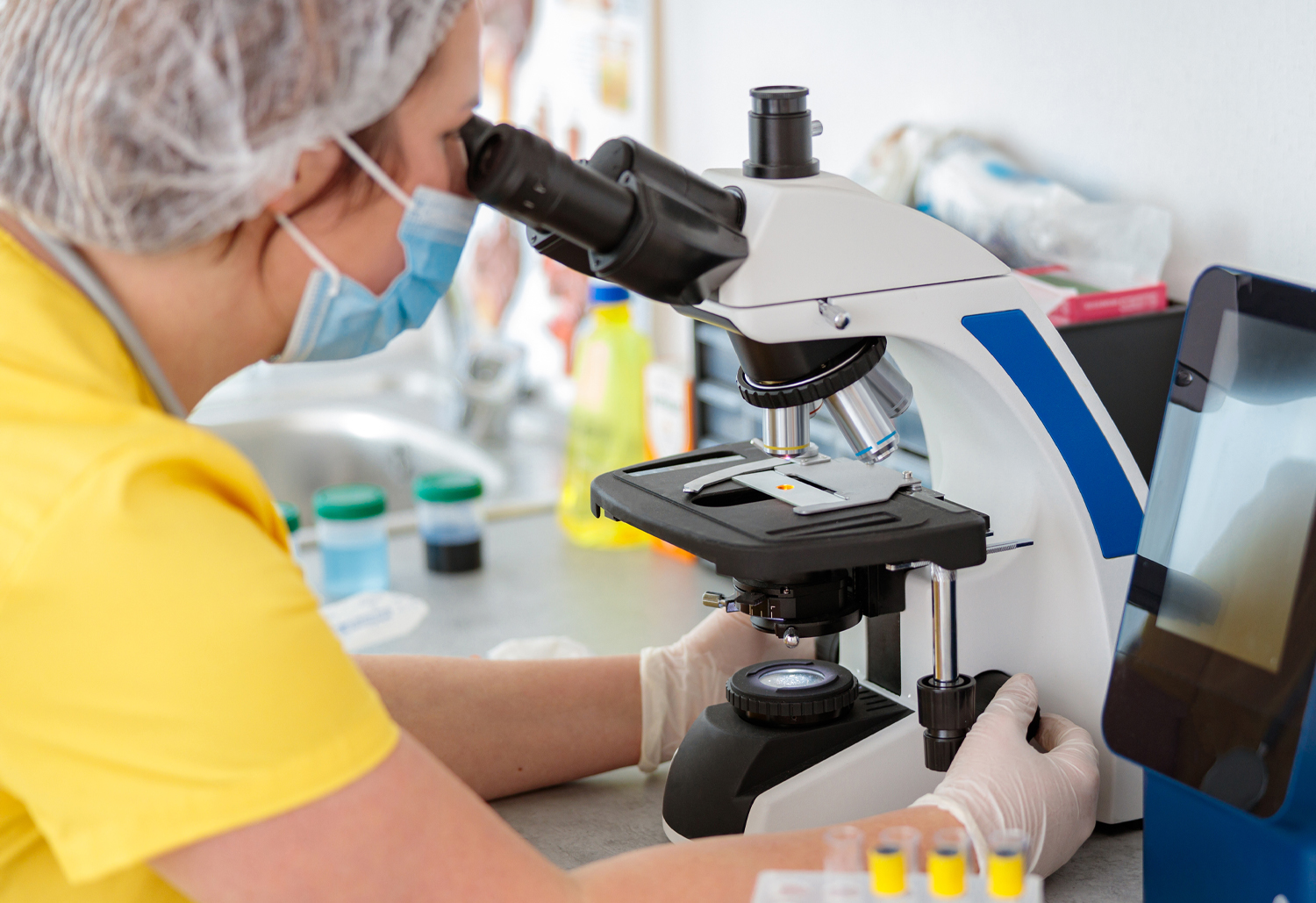
Both cytology and histopathology specimens are stained so that the nucleus, cytoplasm and other structures show up as different colours. The pathologist then examines the slides to come up with a diagnosis.
|